Posterior Muscles Of The Lower Back : Back Muscles Knowledge Amboss - The muscles of the back can be arranged into 3 categories based on their location:
Posterior Muscles Of The Lower Back : Back Muscles Knowledge Amboss - The muscles of the back can be arranged into 3 categories based on their location:. The intermediate layer of back muscles includes the serratus posterior superior and inferior. Related posts of muscles of the lower back and buttocks diagram smooth muscle diagram labeled. Ligaments are tough, flexible bands of connecting tissue that join bones to other bones. Only the iliocostalis is shown on the right side. Aktuelle preise für produkte vergleichen!
The psoas is a hip flexor muscle, as is the quadriceps muscle. The muscles of the lower back help stabilize, rotate, flex, and extend the spinal column, which is a bony tower of 24 vertebrae that gives the body structure and houses the spinal cord. It joins to ligaments and muscles around the pelvis. The extrinsic (superficial) back muscles, which lie most superficially on the back. Pain at the back of the leg which may be sudden onset gradual.
Lower Back Wiktionary from upload.wikimedia.org Muscles also contribute to internal functions of the human body which include motion in the intestines and circulatory system. The psoas is a hip flexor muscle, as is the quadriceps muscle. Muscles of the back complex but divisible into 3 groups (in layers) with different functions: 12 photos of the muscles of the lower back and hip diagram. The pelvic floor muscles also help increase this pressure, which provides stability to the spine and trunk. Here is a depiction of the skeletal frame with the lower. All three subgroups are shown on the left side; The psoas muscle is a low back muscle located deep in the body, very close to the spine and inside the hip and thigh bones.
An extremely strong tendon attached to the heel.
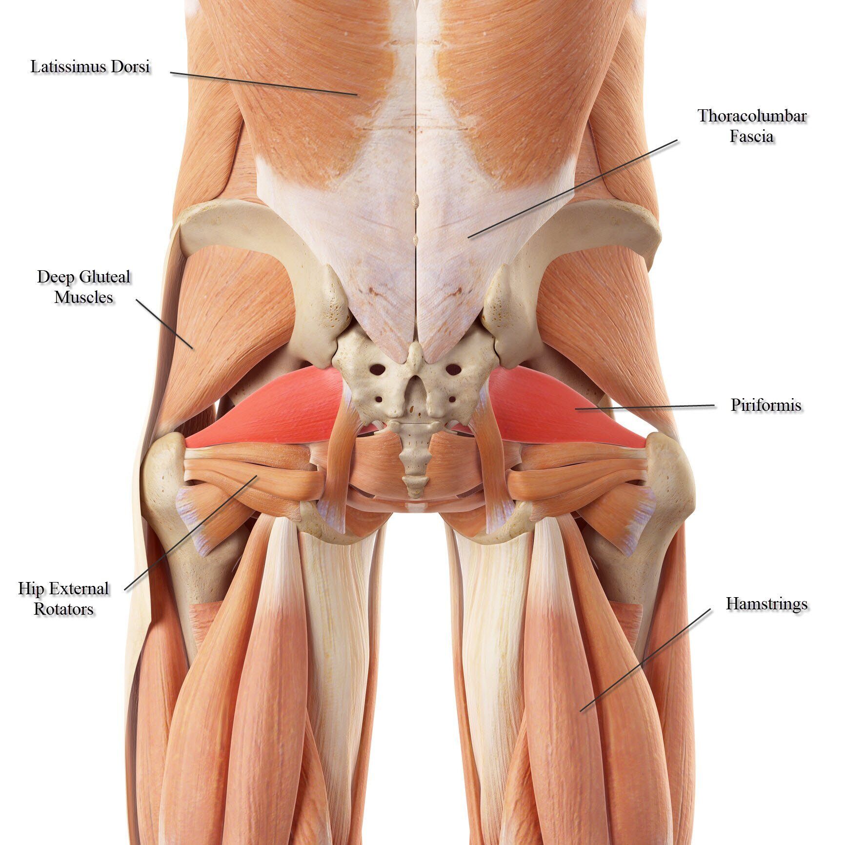
The intermediate layer of back muscles includes the serratus posterior superior and inferior. The butt, including the gluteus minimus, gluteus medius, gluteus maximus The pelvic floor muscles also help increase this pressure, which provides stability to the spine and trunk. The muscles of the back can be arranged into 3 categories based on their location: The psoas is a hip flexor muscle, as is the quadriceps muscle. The erector spinae is composed of three subgroups: These muscles provide posture and stability to the body by holding the vertebral column erect and adjusting the position of the body to maintain balance. It joins to ligaments and muscles around the pelvis. An extremely strong tendon attached to the heel. Two of the main ligaments in the back are the anterior. They help to bend the back to one side or the other. 1 your spine in this region has a natural inward curve. Smooth muscle diagram labeled 12 photos of the smooth muscle diagram labeled labeled diagram of smooth muscle, labeled diagram of smooth muscle cell, smooth muscle cell labeled diagram, smooth muscle diagram labeled, smooth muscle tissue labeled diagram, human muscles, labeled diagram of smooth.
Superficial back muscles, intermediate back muscles and intrinsic back muscles.the intrinsic muscles are named as such because their embryological development begins in the back, oppose to the superficial and intermediate back muscles which develop elsewhere and are therefore classed as extrinsic muscles. Muscles also contribute to internal functions of the human body which include motion in the intestines and circulatory system. These muscles provide posture and stability to the body by holding the vertebral column erect and adjusting the position of the body to maintain balance. The erector spinae is composed of three subgroups: The back is the body region between the neck and the gluteal regions.
Lower Back Muscle Anatomy And Low Back Pain from marvel-b1-cdn.bc0a.com Pain at the back of the leg which may be sudden onset gradual. It joins to ligaments and muscles around the pelvis. Embryonically related to the intercostal muscles, not the deep back mm. An extremely strong tendon attached to the heel. Ligaments are tough, flexible bands of connecting tissue that join bones to other bones. These muscles course from your vertebral column to your ribs and assist your diaphragm and intercostal muscles in moving the ribs during breathing. Smooth muscle diagram labeled 12 photos of the smooth muscle diagram labeled labeled diagram of smooth muscle, labeled diagram of smooth muscle cell, smooth muscle cell labeled diagram, smooth muscle diagram labeled, smooth muscle tissue labeled diagram, human muscles, labeled diagram of smooth. Aktuelle preise für produkte vergleichen!
The muscles of the back are a group of strong, paired muscles that lie on the posterior aspect of the trunk they provide movements of the spine, stability to the trunk, as well as the coordination between the movements of the limbs and the back muscles are divided into two large groups:
The deep muscles develop embryologically in the back, and are thus described as intrinsic muscles. These muscles provide posture and stability to the body by holding the vertebral column erect and adjusting the position of the body to maintain balance. The psoas is a hip flexor muscle, as is the quadriceps muscle. When your lower back feels tight, it's important to listen to your body and take steps to relieve the tension. 12 photos of the muscles of the lower back and hip diagram. The muscles of the lower back help stabilize, rotate, flex, and extend the spinal column, which is a bony tower of 24 vertebrae that gives the body structure and houses the spinal cord. Extrinsic and intrinsic.the back functions are many, such as to house and protect the spinal cord, hold the body and head upright, and adjust the movements of the upper and lower limbs. They also protect the spinal column. 1 your spine in this region has a natural inward curve. Chart of major posterior muscles. The muscles on the back of the trunk help lower the arms and move the body forward and sideways. The intermediate layer of back muscles includes the serratus posterior superior and inferior. Smooth muscle diagram labeled 12 photos of the smooth muscle diagram labeled labeled diagram of smooth muscle, labeled diagram of smooth muscle cell, smooth muscle cell labeled diagram, smooth muscle diagram labeled, smooth muscle tissue labeled diagram, human muscles, labeled diagram of smooth.
The achilles tendon in the strongest in the body. The back is the body region between the neck and the gluteal regions. The erector spinae is composed of three subgroups: All three subgroups are shown on the left side; These muscles provide posture and stability to the body by holding the vertebral column erect and adjusting the position of the body to maintain balance.
Hamstring Muscles And Your Back Pain from www.verywellhealth.com Smooth muscle diagram labeled 12 photos of the smooth muscle diagram labeled labeled diagram of smooth muscle, labeled diagram of smooth muscle cell, smooth muscle cell labeled diagram, smooth muscle diagram labeled, smooth muscle tissue labeled diagram, human muscles, labeled diagram of smooth. Embryonically related to the intercostal muscles, not the deep back mm. Aktuelle preise für produkte vergleichen! The lower spine, the hips and tailbone, and the abdomen. The extrinsic (superficial) back muscles, which lie most superficially on the back. These muscles include the large paired muscles in the lower back, called erector spinae, which help hold up the spine, and gluteal muscles. The muscles on the back of the trunk help lower the arms and move the body forward and sideways. Two of the main ligaments in the back are the anterior.
A respiratory muscle, it receives ventral ramus innervation;
The butt, including the gluteus minimus, gluteus medius, gluteus maximus The latissimus dorsi is a broad, flat muscle that covers the entire lower back. By the way, have you heard about the myth of. It is attached to the calcaneus and is pulled by 3 flexor. Related posts of muscles of the lower back and buttocks diagram smooth muscle diagram labeled. An extremely strong tendon attached to the heel. Since the spine is encircled by musculature, the abdomen, spinal muscles, and hips are all integral aspect of maintaining a healthy lower spine and therefore lower back. The quadratus lumborum muscles (orange, in the image above) are found in the lower back (also called the lumbar area). The lordotic curve your lower back (lumbar spine) is the anatomic region between your lowest rib and the upper part of the buttock. Smooth muscle diagram labeled 12 photos of the smooth muscle diagram labeled labeled diagram of smooth muscle, labeled diagram of smooth muscle cell, smooth muscle cell labeled diagram, smooth muscle diagram labeled, smooth muscle tissue labeled diagram, human muscles, labeled diagram of smooth. A respiratory muscle, it receives ventral ramus innervation; Balance the weight of your head on top of your spine The flexor muscles are attached to the front of the spine and enable flexing, bending forward, lifting, and arching the lower back.
These muscles include the large paired muscles in the lower back, called erector spinae, which help hold up the spine, and gluteal muscles muscles of the lower back. Pain is usually less severe than a hamstring strain although twinges may be felt.


:max_bytes(150000):strip_icc()/GettyImages-87308179-56a05f563df78cafdaa14cd4.jpg)


0 Komentar